Dental X-Rays
Dental X-rays are used to evaluate your oral health. This process uses low levels of radiation to capture images of your teeth and bones. It is a diagnostic tool used to identify dental structures, malignant or benign masses, bone loss and cavities, and dental restorations such as fillings and crowns.
At Oyster Dental, we have RVG (RadioVisioGraphy), which is considered to be one of the best advanced technologies in dentistry. In this, digital radiographs are taken by electronic sensors instead of the traditional system X-ray.
RVG uses significantly lower radiation as compared to that of its traditional counterpart. This makes RVG a safer option for dental images.
Dental X-rays, also known as radiographs, are an indispensable diagnostic tool in modern dentistry. They allow dentists to visualize the internal structures of the teeth, gums, and jawbone, which are not visible during a regular oral examination. Dental X-rays play a crucial role in diagnosing various dental conditions, planning treatments, and monitoring the progression of existing issues.
Types of Dental X-Rays
There are several types of dental X-rays, each serving different diagnostic purposes:
Bitewing X-rays: These X-rays capture the crowns of the upper and lower teeth in a particular section of the mouth. They are commonly used to detect cavities between teeth and to assess bone density supporting the teeth. Bitewing X-rays are taken by having the patient bite down on a small tab attached to the X-ray film.
Periapical X-rays: These provide a detailed view of the entire tooth, from the crown to the root tip and surrounding bone. They are used to detect issues below the gum line, such as abscesses, cysts, and bone loss. Periapical X-rays are particularly useful for diagnosing problems with the tooth’s root and surrounding structures.
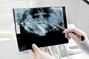
Panoramic X-rays: These X-rays capture a comprehensive view of the entire mouth, including all the teeth, the upper and lower jaws, sinuses, and temporomandibular joints (TMJ). Panoramic X-rays are useful for assessing impacted teeth, jaw disorders, and overall dental and skeletal structures.
Occlusal X-rays: These provide a broad view of the floor of the mouth and the roof of the palate. They are used to detect the position of teeth and their development in children, as well as to identify any abnormalities in the mouth’s structure.
Cone Beam Computed Tomography (CBCT): This advanced imaging technology provides three-dimensional images of the teeth, bones, and soft tissues. CBCT is particularly useful for complex cases, such as implant planning, orthodontic assessments, and evaluating facial structures, as well as identifying the extensions of pathological conditions such as tumors, cysts, fractures of the jaws & planning their treatments. And the structure in association with teeth that are planned for extractions. It’s also used to understand the location of impacted teeth and their associated structures.
Benefits of Dental X-Rays
Dental X-rays offer numerous benefits, including:
Early Detection: X-rays allow dentists to detect dental issues at an early stage, such as cavities, gum disease, and infections. Early detection enables timely intervention, preventing the progression of these conditions and reducing the need for extensive treatments.
Accurate Diagnosis: X-rays provide detailed images of the teeth and supporting structures, helping us to accurately diagnose various Family Dentistry, such as fractures, impacted teeth, and bone loss.
Treatment Planning: Dental X-rays are essential for planning various dental treatments, including orthodontics, root canals, and dental implants. They provide valuable information about the tooth’s structure, root configuration, and bone density.
Monitoring Progress: X-rays help monitor the progress of ongoing treatments, such as orthodontic adjustments, and track the healing process of dental conditions.
Safety Considerations
While dental X-rays involve exposure to low levels of radiation, the benefits far outweigh the risks. Modern dental practices use advanced digital X-ray technology, which significantly reduces radiation exposure. Protective measures, such as lead aprons and thyroid collars, are also used to minimize exposure further.
Patients with concerns about radiation should discuss them with their dentist, who can explain the safety protocols and the necessity of X-rays for their specific dental health needs.
Conclusion
In conclusion, dental X-rays are a vital component of comprehensive dental care. They enable early detection, accurate diagnosis, effective treatment planning, and ongoing monitoring of dental conditions. With advancements in digital imaging technology, it’s safer and more efficient, making them an integral part of maintaining optimal oral health. Regular dental check-ups that include X-rays ensure that potential issues are identified and addressed promptly, contributing to a healthy and confident smile.